Anatomy of a Honey Bee
Anatomy of a Female Honey Bee - Visual
Honey bees are insects and have five characteristics that are common to most insects.
• A hard outer shell called an exoskeleton.
• They have three main body parts: head, thorax, abdomen.
• A pair of antennae that are attached to their head.
• Three pairs of legs used for walking.
• They have two pairs of wings.
You can use the illustrations below to explore the anatomy of the honey bee both what you can see from the outside and also the parts of the honey bee located inside.
Looking at the Outside of a Female Honey Bee
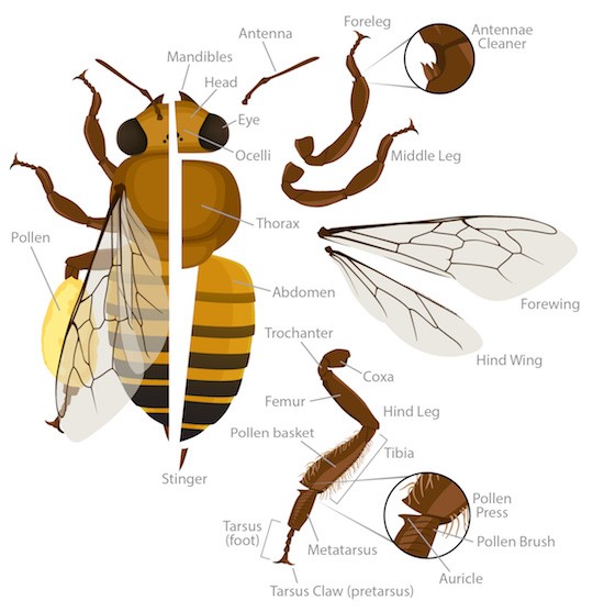
Honey Bee - External
Head | Location of the eyes, brain, where the antennae attach. |
Strong outer mouthparts that help protect the proboscis. | |
(Not shown) Tube-like mouth part used to suck up fluids. | |
One of two types of insect eyes used to detect motion. | |
Eye (Compound) | The second type of eyes made of many light detectors called ommatidia. |
Antenna | Movable segmented feelers that detect airborne scents and currents. |
Midsection where the (6) legs and wings attach. | |
Hind part of the bee and where the stinger is located. | |
Stinger | Or sting, is a sharp organ at the end of the bee’s abdomen used to inject venom. Note: only appears on the female bee. |
Forewings | Wings closest to the head. A bee can fly 15 miles per hour and can travel as much as 3 miles from the hive. The buzzing you hear when you’re near a bee is the rapid flapping of its wings. “They can beat 11,400 times a minute.” |
Hind Wings | Wings farthest from the head. |
Forelegs | Legs closest to the head. |
Antennae Cleaners | Notches filled with stiff hairs that help bees clean their antennae. There is one on each foreleg. |
Middle Legs | Leg located between the foreleg and hind leg. |
Hind Legs | Legs farthest from the head. In workers, these legs have a unique set of tools used to collect and carry pollen called the press, brush, and auricle. |
Coxa | First segment of an insect leg. |
Trochanter | Second segment of an insect leg. |
Femur | Third segment of an insect leg. |
Tibia | Fourth segment of an insect leg; the tibia of the hind leg holds the pollen basket, where pollen is carried. |
Metatarsus | Fifth segment of an insect leg; the metatarsus of the hind leg holds special pollen collecting tools. |
The last segment of the leg and what touches the walking surface. | |
Tarsus Claw | Claw found on the last segment of the leg. The foot of a bee has claws for gripping and a sticky pad for holding on to slippery surfaces. The bee also has taste receptors on its feet. |
Note: Part of the above information has been gathered from: https://www.keepingbackyardbees.com/the-anatomy-of-a-worker-bee/
The Head of a Honey Bee
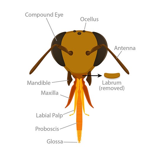
A type of eyes of insect eye that is made of many light detectors called ommatidia. | |
Ocellus | A type of insect eye used to detect motion. (Plural: ocelli) |
Antenna | A movable segmented feeler that detects airborne scents and currents. The bee has 2 antennae which are divided into segments. Female bees have 13 segments where drones have 12. The antennae serves many purposes to the bee. It has over 170 scent receptors. And is used to determine “air speed and orientation during flight.” It is also used to communicate with other bees. “Bees use only the right antennae to communicate.” |
Labrum | Mouthpart that can help handle food and that forms the top of the feeding tube. |
Mandible | Strong outer mouthpart that helps protect the proboscis. |
Maxilla | Mouthpart beneath the mandible that can handle food items. |
Labial Palp | Mouthpart used to feel and taste during feeding. |
Proboscis | Tube-like mouth part used to suck up fluids. |
Glossa | An insect’s hairy tongue that can stick to nectar to pull it in toward the mouth. |
Internal Organs of a Female Honey Bee
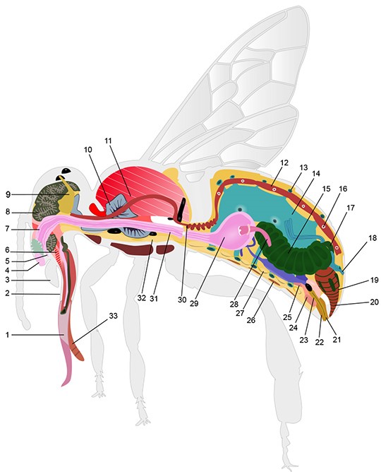
1 | Proboscis | Straw-like mouthparts of a bee used to drink fluids. |
2 | Maxillae | The outer sheath of the proboscis which surrounds the labium. |
3 | Mandible | A pair of jaws used to chew pollen and work wax for comb building. They also help with anything that the bee needs to manipulate. |
4 | Labrum | A movable flap on the head that covers the opening of the food canal and proboscis |
5 | Food Canal | Like our mouths, this is the opening by which the bee will take in food. Bees’ food is almost always liquid in the form of nectar or honey. |
6 | Pharynx | Muscles used to move the labium and suck up nectar from flowers. |
7 | Esophagus | The hollow tube through which ingested fluids pass to the honey stomach and later the midgut. |
8 | Gland that produces some of the compounds necessary for making royal jelly, used to feed the larvae. | |
9 | Brain | Honey bees have excellent learning and memory processing abilities. Their brain processes information used in navigation and communication as well as memory. The brain also controls many of the basic bee body functions. The head of the honey bee holds the brain which is only about a millimeter cubed in size. Though it has a small brain, it has over 950,000 neurons. “The bee brain is one of the most densely packed with neurophil tissue known of any animal.” (see: https://www.keepingbackyardbees.com/the-anatomy-of-a-worker-bee/) |
10 | Salivary Gland | The salivary glands have a number of functions. Like the hypopharyngeal gland, the salivary glands produce some compounds necessary for producing royal jelly. The salivary glands produce liquid used to dissolve sugar, and also produce compounds used to clean the body and contribute to the colony’s chemical identity. |
11 | Flight Muscles | The thorax muscles, which power the bee’s wings for flying and movement. These muscles work very hard and can help the bee to beat its wings up to 230 times per second. |
12 | Heart | Unlike in mammals, honey bees and insects have an open circulatory system, meaning their blood is not contained within tubes like veins or arteries. The blood, or hemolymph, in insects is free-flowing throughout the body cavity and is pumped via the heart. The heart is the structure in red, and acts like a pumping leaky tube to help move the hemolymph throughout the body |
13 | Opening of Spiracle | The respiratory system in insects is a series of hollow tubes connected to air sacs in the body. The openings of these hollow tubes are called spiracles. The tubes are called trachea which then provide oxygen and gas exchange to all tissues in the body. |
14 | Air sac | Air filled sacs used as reservoirs of air in the insect body. |
15 | Midgut | Contains the proventriculus, ventriculus, and small intestine. This is where most of the digestion and nutrient absorption occurs in the insect body |
16 | Heart Openings | Openings in the heart tube which take in and pump out hemolymph. |
17 | Ileum | A short tube connecting the midgut to the hindgut. The Ileum also often houses microbes, which aid in digestion. |
18 | Malpighian Tubules | A set of small tubes that are used to absorb water, waste, and salts and other solutes from body fluid, and remove them from the body. |
19 | Rectum | The rectum acts like our large intestine and is the bees primary location of water absorption for the gut after digestion and nutrient absorption. |
20 | Anus | The exit of the digestive system, used to excrete food waste (poop) while in flight. |
21 | Stinger | Also called "sting" is used to puncture the skin and pump venom into the wound. In worker bees the stinger has a barbed end. Once pushed into the skin the stinger remains in the victim. The venom sac will remain with the stinger. If left in the body the stinger will continue to pump venom from the venom sac into the victim. Queen bees have a longer and un-barbed stinger. Drones (males) do not have a stinger. |
22 | Stinger Sheath | The hardened tube, from which the stinger can slide in and out. |
23 | Sting Canal | The sting is hollow, allowing venom to pass through the stinger. This is also the canal via which an egg is passed, when the queen lays an egg. |
24 | Venom Sack | Holds the venom produced by the venom gland, and can then contract to pump venom through the stinger. |
25 | Venom Gland | The gland which produces the venom that damages tissue if injected into the body. |
26 | Worker bees start to secrete wax about 12 days after emerging. About six days later the gland degenerates and that bee will no longer produce wax. The queen is continually laying eggs to maintain colony size and to produce more new workers that produce wax. | |
27 | Ventral Nerve Cord | Like the nerve cord in our spine, which holds bundles of nerve fibers that sends signals from our brain to the rest of our body. |
28 | Proventriculus | A constricted portion of the honey bee foregut or honey stomach, which can control the flow of nectar and solids. This allows honey bees to store nectar in the honey stomach without being digested. |
29 | Honey Stomach (Foregut/Crop) | A storage sac, used in honey bees to carry nectar. The honey stomach is hardened to prevent fluids from entering the body at this location. |
30 | Aorta | Blood vessel located in the back of a bee that carries blood from the heart to the organs. |
31 | Esophagus | Part of the bee digestive system that begins below the mouth and connects to the honey stomach. |
32 | Ventral Nerve Cord | Same as 27. This is a large bundle of nerves from the brain that sends signals to the rest of the bee’s body. |
33 | Labium | In bees a tongue-like appendage used to help drink up nectar. Like our tongue bees can taste with this organ. The labium fits inside of the maxilla (2), kind of like a straw. |
Citation: Christopher M. Jernigan. (2017, June 13). Bee Anatomy. ASU – Ask A Biologist. Retrieved September 10, 2020 from https://askabiologist.asu.edu/honey-bee-anatomy
The Anatomy of a Worker Bee